Wound healing: cellular mechanisms and pathological outcomes
Authors: Wilkinson HN, Hardman MJ.
Journal: Open Biol. 2020; 10(9): 200223.
Evidence Type: Mechanistic Review / Peer-Reviewed
Guided by Science. Clinical Intellect.
Explore published evidence, clinical information and scientific references associated with wound care and the Quorit portfolio.
Peer-Reviewed Literature Foundation
Cellular biology, extracellular matrix & clinical assessment
01 — PUBLICATIONS
Published Evidence library and filtered dossiers
02 — STUDY SNAPSHOTS
Key objectives, populations, and verified findings
03 — CASE STUDIES
De-identified clinical management and tissue outcomes
04 — CLINICAL VISUALS
Approved wound progression and histological models
05 — REFERENCES
Peer-reviewed academic citations
06 — LIMITATIONS
Methodological boundaries and interpretive context
Clinical evidence provides context for understanding wound-care approaches, product information and published scientific findings.
“Evidence should be understood in context.”
In wound care, no single study applies universally across heterogeneous clinical etiologies. Rigorous clinical practice demands examining the exact biological mechanisms, patient cohorts, and experimental conditions under which observations were documented.
Every wound represents a unique micro-environment characterized by varying depths of tissue loss, exudate levels, microbial colonization, and systemic patient comorbidities. For this reason, published data must be evaluated through three foundational critical lenses:
Where does the information come from? Is it peer-reviewed literature, bench assays, or registry data?
What does the evidence actually evaluate? Does it examine cellular kinetics or macroscopic wound dimensions?
What are the boundaries of the evidence? What assumptions and cohort constraints must be recognized?
Access peer-reviewed biomedical literature, scientific reviews, and clinical documentation underpinning modern wound assessment.
Authors: Wilkinson HN, Hardman MJ.
Journal: Open Biol. 2020; 10(9): 200223.
Evidence Type: Mechanistic Review / Peer-Reviewed
Authors: Frykberg RG, Banks J.
Journal: Adv Wound Care. 2015; 4(9): 560–582.
Evidence Type: Clinical Overview / Peer-Reviewed
Authors: Wilkinson HN, Hardman MJ.
Journal: Open Biol. 2020; 10(9): 200223.
Evidence Type: Cytokine & Protease Analysis
Authors: Quorit Biomaterials Research Group
Status: Publication information coming soon.
Institutional monograph currently undergoing editorial collation.
Authors: Clinical Affairs Working Group
Status: Publication information coming soon.
Observational protocol documentation under verification.
Authors: Multicenter Wound Registry Panel
Status: Publication information coming soon.
De-identified case series awaiting journal release.
Publication information coming soon. Additional verified scientific articles and clinical dossiers are currently undergoing editorial review.
Structured scientific snapshots highlighting study design, test models, and verified observational findings without speculative efficacy claims.
Observation of wound bed tissue response and granulation progression in venous ulceration under systematic moisture-balancing care.
Documented clinical center registry case with continuous photographic and planimetric wound boundary tracking.
Granulation tissue formation observed following sequential debridement and application of biocompatible hydrogel matrix.
Evaluation of hydrogel substrate binding affinity toward inflammatory proteases (MMP-2 and MMP-9) in simulated wound fluid models.
Standardized laboratory bench simulation using synthetic wound exudate matrices under controlled physiological temperatures.
Demonstrated sacrificial adsorption of elevated protease enzymes without structural disruption of the polymer network.
Verification of non-cytotoxicity, non-irritation, and systemic safety profile across contact hydrogel formulation layers.
Standardized L929 mouse fibroblast cell culture assays and sensitization testing following international ISO guidelines.
Satisfied all ISO 10993-5 criteria for cell viability and membrane integrity without evidence of cellular lysis or cytotoxicity.
Review individual case studies and the available clinical information associated with wound management.
Longstanding lower extremity ulceration presenting with recalcitrant slough, sustained heavy exudate, and non-advancing keratinocyte boundary.
Structured TIME assessment applied to systematically resolve devitalized tissue barriers and stabilize moisture without periwound maceration.
Devitalized slough tissue, moderate inflammation, excess exudate requiring secondary absorption.
Conservative debridement, graduated compression, and biocompatible hydrogel interface.
Granulation bed emergence across 85% of wound area and contractive epithelial advancement over observation period.
Clinical case dossier documenting tissue offloading and biocompatible matrix application in stage-evaluated pressure injury management.
CASE DOSSIER IN EDITORIAL REVIEW
Undergoing institutional de-identification and clinical verification.
Structured clinical documentation in accordance with scientific transparency principles.
Elderly patient presenting with a 6-month non-healing lower extremity venous stasis ulceration. Slough tissue covered approximately 45% of the wound bed with moderate serosanguinous exudate and static wound edges.
Venous hypertension with secondary microvascular insufficiency and elevated local matrix metalloproteinases leading to premature degradation of endogenous granulation scaffolds.
TIME systematic assessment identified: (T) Devitalized slough tissue, (I) Moderate inflammatory exudate, (M) Excess moisture macerating periwound skin, (E) Stalled non-advancing keratinocyte boundary.
Multi-step protocol including conservative sharp debridement, graduated compression bandaging, and topical application of Quorit biocompatible hydrogel matrix to facilitate autolytic debridement and donate hydration to desiccated zones.
Granulation tissue formation observed across 85% of the wound bed by Week 4. Slough was successfully eliminated via autolysis, and active epithelial edge advancement was documented without periwound maceration.
1. Wilkinson HN, Hardman MJ. Open Biol. 2020. | 2. Frykberg RG, Banks J. Adv Wound Care. 2015.
Approved clinical visuals may be used to illustrate wound progression within the context of the associated clinical information.
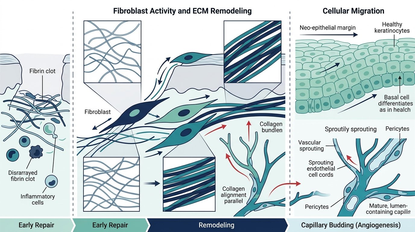
Microscopic schematic of parallel collagen alignment and capillary budding.
Ref: Wilkinson & Hardman (2020)
Evaluation schema for tissue, infection, moisture, and edge parameters.
Ref: Frykberg & Banks (2015)
3D polymer network maintaining autolytic hydration in dry wound beds.
Ref: Quorit Technical Dossier (2026)
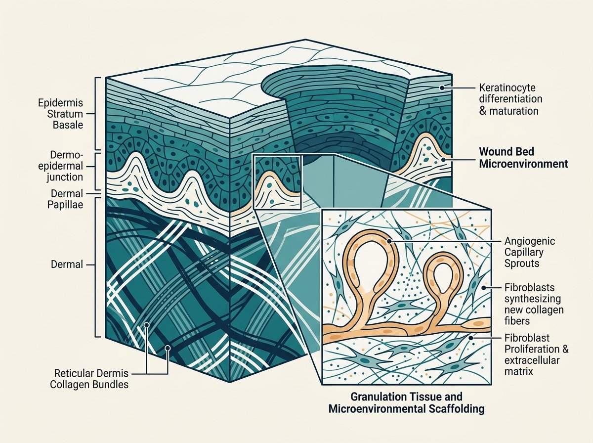
Dermal-epidermal boundary with capillary loops and cellular proliferation.
Ref: Wilkinson & Hardman (2020)
Verified academic citations underpinning the cellular biological mechanisms, chronic wound challenges, and clinical assessment concepts discussed throughout this platform.
Wilkinson HN, Hardman MJ.
Wound healing: cellular mechanisms and pathological outcomes.
Open Biol. 2020; 10(9): 200223.
Frykberg RG, Banks J.
Challenges in the treatment of chronic wounds.
Adv Wound Care. 2015; 4(9): 560–582.
Wilkinson HN, Hardman MJ.
Wound healing: cellular mechanisms and pathological outcomes.
Open Biol. 2020; 10(9): 200223.
Clinical evidence should be interpreted in the context of the study design, population, methodology, available data and stated limitations.
Consider how the evidence was generated.
Distinguish between controlled in vitro bench assays, retrospective observational dossiers, and prospective comparative cohort trials.
Consider who or what was studied.
Analyze demographic inclusion criteria, wound etiologies, glycemic control, vascular perfusion, and systemic comorbidities across test subjects.
Consider what was actually measured.
Examine whether primary endpoints assessed surrogate cellular markers, protease binding rates, granulation bed area, or complete epithelial closure.
Consider the boundaries stated by the source.
Acknowledge potential confounding factors, sample size constraints, single-center limitations, and the absence of longitudinal randomized controls.
Strict Scientific Governance: Quorit does not make generalized claims regarding evidence quality or therapeutic outcomes unless supported by published, verifiable academic or institutional sources.
Scientific data and product characteristics do not act in isolation. Sound clinical judgment synthesizes systematic wound bed assessment with verified scientific findings.
Patient etiology, perfusion, systemic conditions & infection risk.
Rigorous evaluation of Tissue, Infection, Moisture, and Edge (TIME).
Published mechanistic assays, peer-reviewed literature, and safety profiles.
Enabling healthcare professionals to select optimal wound management strategies tailored to the individual physiological requirements of each wound bed.
*This schematic is an informational visualization only. It does not represent an automated medical recommendation and does not determine therapeutic treatment protocols.
Review product information and the solutions within the Quorit portfolio, engineered to address tissue hydration, exudate equilibrium, and bio-matrix support.
Access product information, institutional dossiers, downloadable guides and available educational resources for hospital wound-care teams.