TIME:
A Systematic Approach to Wound Assessment
How Do We Know What Is Holding the Wound Back?
A systematic wound assessment helps turn a complex wound into identifiable clinical problems.
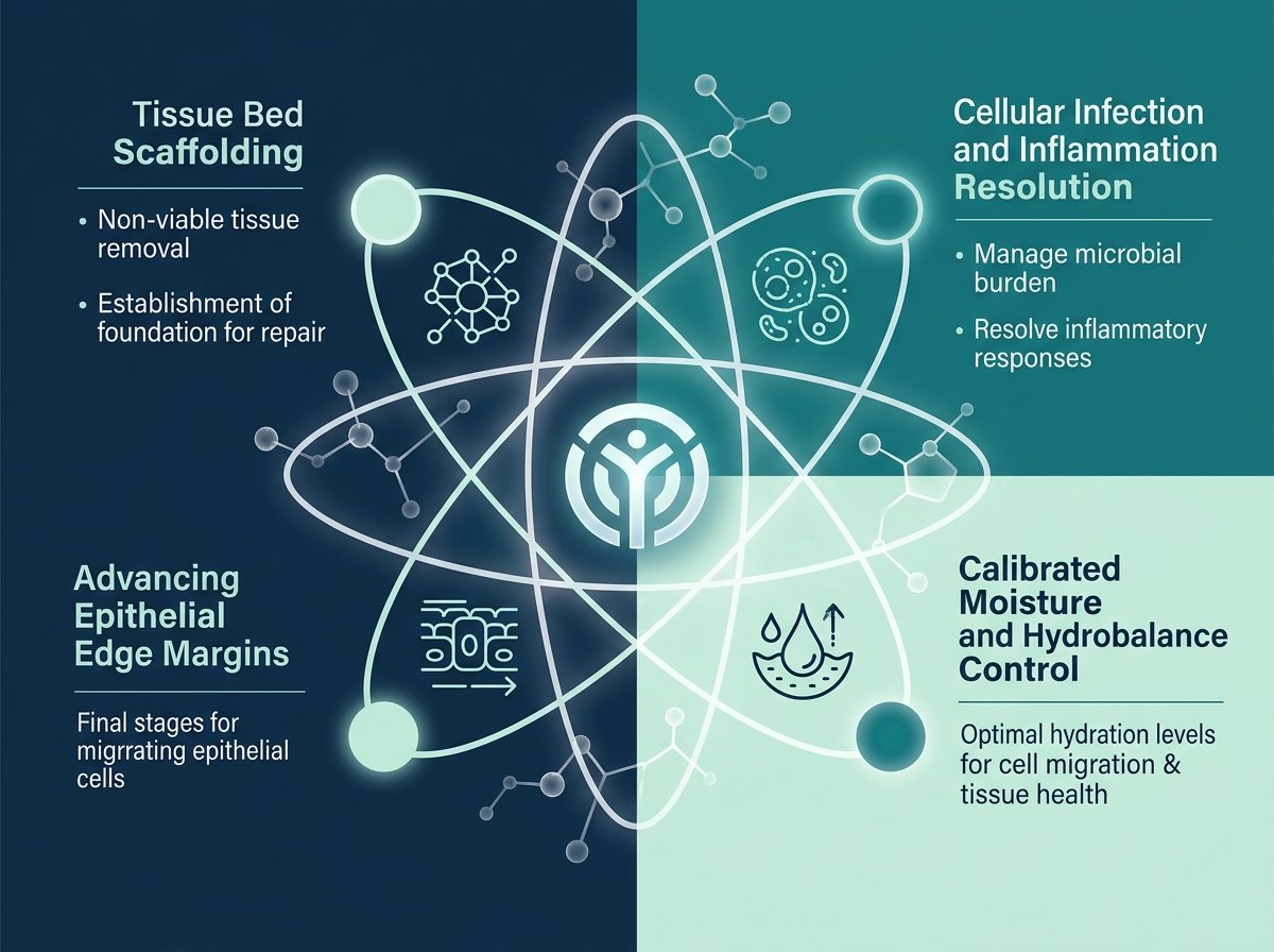
The TIME Approach
TISSUE
Identify and remove non-viable or deficient tissue to establish a vascularized bed.
INFECTION / INFLAMMATION
Control persistent bioburden, biofilms, and excessive destructive protease activity.
MOISTURE
Calibrate optimal hydrobalance to prevent peri-wound maceration or tissue desiccation.
EDGE
Promote centripetal keratinocyte advancement and overcome undermining barriers.
What Is in the Wound Bed?
- Necrotic tissue / eschar
- Slough
- Deficient tissue
“Is the wound bed ready to heal?”
Remove barriers and create a viable wound bed.
Different types of debridement are used to restore a viable wound environment.
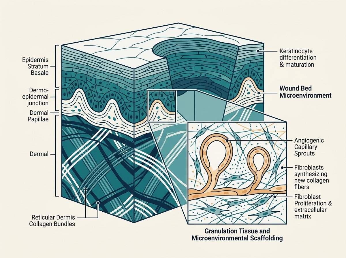
NECROTIC TISSUE / ESCHAR
NON-VIABLE BARRIERBlack, brown, or hardened devitalized tissue adhering firmly to the wound base or margins. Acts as a physical and biological barrier preventing granulation capillary formation, re-epithelialization, and cellular penetration.
Is the Wound Still in Defense Mode?
Persistent cellular activation and sustained microbial bioburden prevent progression from inflammation to the proliferative phase.
“Is inflammation resolving—
or is something keeping it active?”
Identify and address infection, excessive inflammation and microbial factors that may be delaying healing.
Local, systemic or intravenous antimicrobial agents are needed as per clinical features and bacterial bioburden.
Microscopic Equilibrium
Transition toward M2 anti-inflammatory macrophage phenotypes. Down-regulation of active MMPs, allowing newly synthesized fibronectin and type-I collagen fibril assembly.
Sustained bacterial biofilm colonization and neutrophil extracellular trap release. Continuous enzymatic cleavage of essential growth factor receptors keeps tissue in sterile defense.
PERSISTENT INFLAMMATION
LOCAL / SYSTEMIC SIGNS OF INFECTION
INCREASING PAIN OR DETERIORATION
MALODOR / PURULENT EXUDATE
MICROBIAL BURDEN / BIOFILM
Is the Wound Environment Balanced?
Optimal healing occurs within a calibrated moisture window. Both excessive exudate and excessive dryness compromise cellular viability.
Moisture Continuum
Hover or click any state to calibrate assessment position:
Supports cellular signaling, facilitates enzymatic autolysis of fibrinous residues, and provides an ideal fluid medium for growth factors, nutrient delivery, and cell mitosis.
✓ supports healing & cell viabilityAmount of Exudate
None, low, moderate, heavy, or copious.Character of Exudate
Serous, haemoserous, sanguineous, purulent.Maceration
Softened, waterlogged, or white peri-wound margins.Excess Dryness
Crusting, scabbing, or desiccation stalling migration.Peri-Wound Condition
Erythema, excoriation, or fungal satellite lesions.“Does the wound have the right environment for repair?”
With exudate management (Foam dressing) and protection of the wound edge being central considerations.
Standard clinical rationale prioritizes dressings that absorb excess fluid while maintaining a moist wound interface.
Is the Wound Actually Moving Toward Closure?
Assessment of epithelial migration margins and surrounding skin integrity to determine whether centripetal closure is progressing.
Centripetal migration of pink epithelial tissue toward center.
Healthy Centripetal Migration
The epidermal margins are flush with the wound bed, moist, and exhibiting a delicate translucent silvery-pink border representing active keratinocyte mitosis and crawling across a viable collagen matrix.
Rolled, epibole, or undermined edge stalling migration.
Epibole & Undermining Barriers
Keratinocytes reach the margin and roll downward instead of migrating outward because the wound bed lacks viable scaffolding. Undermining creates dead-space pockets harboring bioburden.
- Advancing epithelial edge
- Non-advancing edge
- Undermining
- Condition of surrounding skin
“Is new skin moving across the wound—
or has progress stalled?”
Identify barriers preventing epithelial advancement and wound closure.
E around non-advancing or undermined wound edges; later descriptions also emphasize the surrounding skin.
TIME Turns Complexity Into Clinical Questions.
A standardized, peer-reviewed framework aligning observation with actionable intervention.
TISSUE
“What is in the wound bed?”
INFECTION / INFLAMMATION
“Is the wound still in defense mode?”
MOISTURE
“Is the wound environment balanced?”
EDGE
“Is the wound moving toward closure?”
From Assessment to Action
What Did We Find?
| ASSESSMENT | WHAT MAY BE PREVENTING PROGRESS? |
|---|---|
| T T — Tissue | Non-viable / deficient tissue |
| I I — Infection / Inflammation | Persistent inflammation / microbial burden |
| M M — Moisture | Too dry / too wet / uncontrolled exudate |
| E E — Edge | Non-advancing / undermined wound edge |
Non-viable / deficient tissue
Persistent inflammation / microbial burden
Too dry / too wet / uncontrolled exudate
Non-advancing / undermined wound edge
Ongoing Clinical Process
A dynamic loop continuously adjusting to wound progression:
ASSESS
Systematic TIME evaluation of baseline wound status.
IDENTIFY
Pinpoint exact barriers hindering progression.
ADDRESS
Deploy tailored therapeutic wound-care solutions.
REASSESS
Monitor healing trajectory and iterate clinical strategy.
From Assessment
to Appropriate Solutions
Explore the Quorit portfolio after understanding the wound and considering the key factors that may influence its progression.
SCIENTIFIC REFERENCES
Literature supporting structured tissue viability assessment and wound microenvironmental management:
Wilkinson HN, Hardman MJ. Wound healing: cellular mechanisms and pathological outcomes. Open Biol. 2020.
Frykberg RG, Banks J. Challenges in the treatment of chronic wounds. Adv Wound Care. 2015;4(9):560-582.
Wilkinson HN, Hardman MJ. Wound healing: cellular mechanisms and pathological outcomes. Open Biol. 2020.